Home » Without Label » Pelvic Ultrasound Female : ultrasound of Female Pelvic Inflammatory Disease - YouTube / Female pelvis ultrasound sonography plays the primary role in imaging of the female pelvis.
Pelvic Ultrasound Female : ultrasound of Female Pelvic Inflammatory Disease - YouTube / Female pelvis ultrasound sonography plays the primary role in imaging of the female pelvis.
Pelvic Ultrasound Female : ultrasound of Female Pelvic Inflammatory Disease - YouTube / Female pelvis ultrasound sonography plays the primary role in imaging of the female pelvis.. Doppler ultrasound of the female pelvis. Excretory organs like the kidneys, urethra, bladder and bowel, and surrounding. Doppler ultrasound of the female pelvis. However, it is considered more invasive than the transabdominal approach. General uses in both men and women include evaluating bladder problems, bladder tumors, kidney stones, and pelvic pain and masses.
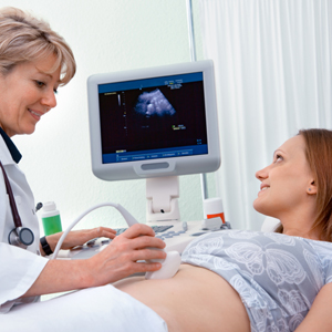
Ultrasound imaging uses soundwaves to create pictures of the inside of the body. A transvaginal ultrasound, also called an endovaginal ultrasound, is a type of pelvic ultrasound used by doctors to examine female reproductive organs. The sound waves create a picture on a video monitor. Ultrasound imaging of the pelvis uses sound waves to produce pictures of the structures and organs in the lower abdomen and pelvis. A pelvic ultrasound is a test that uses sound waves to make pictures of the organs inside your pelvis.
Women's Private Pelvic Scan - Fertility + Bloods ... from www.ultrasound-direct.com A female pelvis ultrasound, a.k.a. A pelvic ultrasound is a test that uses sound waves to make a picture of the inside of the lower belly (pelvis). General uses in both men and women include evaluating bladder problems, bladder tumors, kidney stones, and pelvic pain and masses. Doppler ultrasound of the female pelvis. A transvaginal ultrasound, or endovaginal ultrasound, is a safe and straightforward procedure that doctors use to examine the internal organs in the female pelvic region. This has been recognised by bodies such as the royal college of obstetricians and gynaecologists, who have made ultrasound part of the core requirements in their. Your doctor might order this test to diagnose a condition, or to check the health of your baby. Watch this and know what to expect before your appointment.
In children, pelvic ultrasounds can give doctors more information in cases of ambiguous genitalia.
A transvaginal ultrasound, also called an endovaginal ultrasound, is a type of pelvic ultrasound used by doctors to examine female reproductive organs. Ultrasound can distinguish whether a uterine polyp is causing the bleeding you are experiencing in between your normal cycles. Ultrasound is integral to modern gynaecological practice. An ultrasound may help your health care provider figure out the cause of your pain, lack of periods, or irregular periods, the position of an iud or if you are pregnant. This includes the uterus, fallopian tubes,. General uses in both men and women include evaluating bladder problems, bladder tumors, kidney stones, and pelvic pain and masses. A pelvic ultrasound is a test that uses sound waves to make a picture of the inside of the lower belly (pelvis). However, it is considered more invasive than the transabdominal approach. A pelvic ultrasound can be performed at any stage of a woman's menstrual cycle. This has been recognised by bodies such as the royal college of obstetricians and gynaecologists, who have made ultrasound part of the core requirements in their. Ultrasound uses a transducer that sends out ultrasound waves at a frequency too high to be heard. If you are pregnant, these exams are common. Doppler ultrasound of the female pelvis.
However, it is considered more invasive than the transabdominal approach. Summary this review provides updates of recent changes in female pelvic ultrasound imaging, and we hope it will aid radiologists in accurate diagnosis and management of gynecologic diseases. A pelvic ultrasound allows quick visualization of the female pelvic organs and structures including the uterus, cervix, vagina, fallopian tubes and ovaries. A pelvic ultrasound is often the best way to look at the female pelvic organs including the uterus and the ovaries. If you are pregnant, these exams are common.
Pelvic Ultrasound | Center for Young Women's Health from youngwomenshealth.org A pelvic ultrasound is a test that uses sound waves to make a picture of the inside of the lower belly (pelvis). This includes the uterus, fallopian tubes,. An ultrasound of the pelvis is typically used to look at the bladder, ovaries, uterus, cervix, and fallopian tubes (some of these are known as the female reproductive organs). Female pelvic ultrasound procedure is for indicating the status of endometrium, ovaries and uterus. General uses in both men and women include evaluating bladder problems, bladder tumors, kidney stones, and pelvic pain and masses. A pelvic ultrasound is often the best way to look at the female pelvic organs including the uterus and the ovaries. The purpose of a pelvic ultrasound can depend on whether you are male or female. An ultrasound may help your health care provider figure out the cause of your pain, lack of periods, or irregular periods, the position of an iud or if you are pregnant.
A pelvic ultrasound can be performed at any stage of a woman's menstrual cycle.
Some women experience changes in their menstrual cycle in length or quantity. However, it is considered more invasive than the transabdominal approach. Ultrasound uses a transducer that sends out ultrasound waves at a frequency too high to be heard. A pelvic ultrasound allows quick visualization of the female pelvic organs and structures including the uterus, cervix, vagina, fallopian tubes and ovaries. Excretory organs like the kidneys, urethra, bladder and bowel, and surrounding. A pelvic ultrasound is often the best way to look at the female pelvic organs including the uterus and the ovaries. An ultrasound may help your health care provider figure out the cause of your pain, lack of periods, or irregular periods, the position of an iud or if you are pregnant. A transvaginal ultrasound, also called an endovaginal ultrasound, is a type of pelvic ultrasound used by doctors to examine female reproductive organs. This includes the uterus, fallopian tubes,. A hand held device called a transducer (also called a probe or wand) sends and receives these soundwaves. Like an annual pelvic exam, a transvaginal ultrasound is typically performed with the patient lying on her back on the exam table with her feet in stirrups. Female pelvic ultrasound phantom (transvaginal) the pelvic phantom is unique in that it allows for transabdominal and transvaginal scanning. If you are pregnant, these exams are common.
A pelvic ultrasound allows quick visualization of the female pelvic organs and structures including the uterus, cervix, vagina, fallopian tubes and ovaries. The test can be done in two ways: Primary indications for female pelvic us examination are pelvic pain, abnormal vaginal bleeding, and suspicion of pelvic mass. The sound waves create a picture on a video monitor. Though transvaginal scanning is the gold standard for gyn and 1st trimester pregnancy, it is still important to recognize the anatomy from the transabdominal aspect, as patients may present with anatomy and/or pathology that is outside the transvaginal.
Normal transabdominal pelvic ultrasound | Image ... from images.radiopaedia.org In children, pelvic ultrasounds can give doctors more information in cases of ambiguous genitalia. A transvaginal ultrasound, or endovaginal ultrasound, is a safe and straightforward procedure that doctors use to examine the internal organs in the female pelvic region. A pelvic ultrasound allows quick visualization of the female pelvic organs and structures including the uterus, cervix, vagina, fallopian tubes and ovaries. An ultrasound may help your health care provider figure out the cause of your pain, lack of periods, or irregular periods, the position of an iud or if you are pregnant. The test can be done in two ways: Ultrasound is integral to modern gynaecological practice. This includes the uterus, fallopian tubes,. An ultrasound of the pelvis is typically used to look at the bladder, ovaries, uterus, cervix, and fallopian tubes (some of these are known as the female reproductive organs).
This has been recognised by bodies such as the royal college of obstetricians and gynaecologists, who have made ultrasound part of the core requirements in their.
A hand held device called a transducer (also called a probe or wand) sends and receives these soundwaves. Ultrasound imaging of the pelvis uses sound waves to produce pictures of the structures and organs in the lower abdomen and pelvis. A transvaginal ultrasound, also called an endovaginal ultrasound, is a type of pelvic ultrasound used by doctors to examine female reproductive organs. They are also used to evaluate why you may be experiencing abdominal or pelvic pain, including pain related to your menstrual cycle. If you are pregnant, these exams are common. The sound waves create a picture on a video monitor. There are three types of pelvic ultrasound: Excretory organs like the kidneys, urethra, bladder and bowel, and surrounding. Ct and mr are supplemental techniques used when the us examination is equivocal and in the staging of pelvic malignancy. A pelvic ultrasound can be performed at any stage of a woman's menstrual cycle. General uses in both men and women include evaluating bladder problems, bladder tumors, kidney stones, and pelvic pain and masses. Complete pelvic ultrasound (upeltv) this is a complete pelvic ultrasound exam, including transabdominal and transvaginal. A pelvic ultrasound allows quick visualization of the female pelvic organs and structures including the uterus, cervix, vagina, fallopian tubes and ovaries.